Thyroid Eye Disease Affects 16 in 100,000 Women Yearly, Smoking Multiplies Risk
The U.S. Food and Drug Administration approved teprotumumab-trbw in January 2020 for the treatment of Graves’ ophthalmopathy, also known as thyroid eye disease. This autoimmune condition occurs in 16 per 100,000 women annually and 3 per 100,000 men, according to medical sources.
Prevalence and Risk Factors
Thyroid eye disease most commonly affects individuals with Graves’ disease, though it can also occur in those with Hashimoto’s thyroiditis or who are euthyroid. Cigarette smoking raises the incidence of the disease 7.7-fold, significantly increasing risk for smokers. About 3 to 5 percent of cases involve severe disease marked by intense pain, sight-threatening corneal ulceration, or compression of the optic nerve, which can threaten vision.
Symptoms and Disease Progression
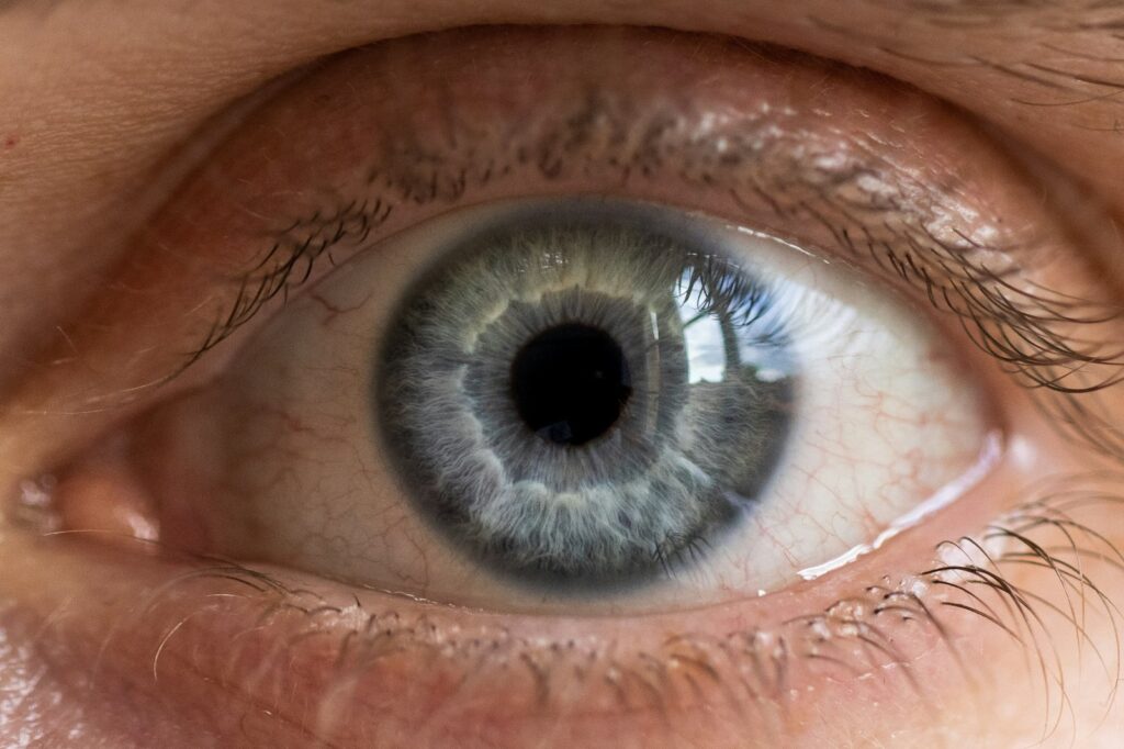
In mild disease, patients often experience eyelid retraction, lid lag, swelling, redness, conjunctivitis, and bulging eyes. Non-specific symptoms include irritation, grittiness, photophobia, tearing, and blurred vision. Pain is not typical, but pressure in the orbit is commonly reported. In moderate to severe active disease, inflammation and edema of extraocular muscles lead to gaze abnormalities, double vision, and restrictive myopathy. Severe cases may involve optic neuropathy, progressive exophthalmos, and loss of visual acuity, requiring urgent intervention to prevent permanent blindness.
Treatment Evolution and Future Outlook
Treatment has evolved from reliance on glucocorticoids and surgical options to targeted therapies. Teprotumumab-trbw (Tepezza; Amgen) and veligrotug-vvze (Lumvoa; Viridian Therapeutics) are insulin-like growth factor 1 receptor antagonists that have helped patients avoid surgery or high-dose systemic corticosteroids. More subcutaneous and oral targeted therapies are in development. At the American Academy of Ophthalmology 2026 Annual Meeting, experts emphasized the importance of reviewing imaging personally and noted that artificial intelligence may assist in screening orbital imaging and determining whether scans are normal or abnormal. Patients should consult their doctor about appropriate care options. Early findings suggest that advances in treatment and diagnostics are improving outcomes, and the encouraging part is that more options now exist beyond steroids or surgery. Talk to your doctor to understand what is right for your situation. Consult your doctor.
Diagnostic Imaging Guidelines and Clinical Recommendations
Clinicians should image most, if not all, patients with thyroid eye disease at initial presentation, especially when the diagnosis is uncertain or there are atypical features. Computed tomography is quicker than magnetic resonance imaging and should be performed without contrast to avoid the risk of worsening active disease with iodine contrast. CT is also useful for patients with significant proptosis who may need bony orbital decompression, as it better images bony anatomy.
Magnetic resonance imaging is preferred when another diagnosis, such as a tumor or other orbital inflammation, is in question, because it better images soft tissue. In cases of dysthyroid optic neuropathy, both CT and MRI are often obtained, as these patients frequently need decompression and MRI helps assess optic nerve inflammation. The key pitfall to avoid is relying solely on the radiology report.
Clinicians should always review the images themselves, especially if there are subtle findings that radiologists may not pick up. This practice ensures accurate interpretation and supports informed clinical decision-making in the management of thyroid eye disease.
Treatment Evolution from Steroids to Targeted Therapies
Historically, severe thyroid eye disease was managed with glucocorticoids (steroids) and sometimes ciclosporin, as noted in the source material. Many anti-inflammatory biological mediators, such as infliximab, etanercept, and anakinra, were being tried for treatment. This approach aimed to reduce inflammation and manage symptoms before the advent of targeted therapies.
In January 2020, the U.S. Food and Drug Administration approved teprotumumab-trbw for the treatment of Graves’ ophthalmopathy, marking a significant shift in treatment strategy. This insulin-like growth factor 1 receptor antagonist (Tepezza; Amgen) represented the first targeted therapy approved specifically for thyroid eye disease, offering an alternative to high-dose systemic corticosteroids and surgery.
More recently, veligrotug-vvze (Lumvoa; Viridian Therapeutics) has emerged as another insulin-like growth factor 1 receptor antagonist targeting the same pathway. Both therapies have helped patients avoid surgery or reduce reliance on high-dose corticosteroids. The development of additional subcutaneous and oral targeted therapies is ongoing, expanding options beyond traditional steroid-based regimens.
Impact on Daily Functioning and Quality of Life
Thyroid eye disease can significantly affect daily activities and overall quality of life, particularly due to visual disturbances and ocular discomfort. Patients with moderate to severe disease often experience double vision, which may interfere with reading, driving, or performing tasks requiring binocular vision. The restriction of eye movements from fibrosis of the extraocular muscles can lead to gaze abnormalities, making it difficult to look in certain directions without head movement.
Symptoms such as irritation, grittiness, photophobia, tearing, and blurred vision are commonly reported, even in mild cases, contributing to chronic discomfort. Eyelid retraction and lagophthalmos increase the risk of corneal exposure and dryness, necessitating frequent use of artificial tears or lubricants. In severe cases, optic neuropathy and progressive exophthalmos may result in permanent visual field loss or color vision deficits if not treated urgently.
The psychosocial impact is also notable, as visible changes like bulging eyes and eyelid retraction can affect self-esteem and social interactions. While mild disease may resolve with supportive measures such as artificial tears and smoking cessation, active inflammation often requires medical intervention to prevent functional impairment and preserve both vision and quality of life. Early diagnosis and personalized treatment are essential to minimize long-term disability.
Artificial Intelligence as a Screening Tool for Orbital Imaging
During the AAO 2026 meeting, Dr. Liza M. Cohen explained that machine‑learning platforms are poised to assist clinicians by automatically sorting orbital scans into “normal” versus “potentially abnormal” categories. This triage function would let physicians focus their attention on studies most likely to require intervention.
Cohen also indicated that algorithmic output could contribute to forming an initial diagnostic impression, helping to guide the ophthalmologist’s assessment rather than replacing expert interpretation. The anticipated workflow mirrors the broader move toward computational support in thyroid eye disease evaluation.
She emphasized that, even with AI assistance, clinicians must still personally review images—especially subtle abnormalities that radiology reports might overlook—to ensure accurate decision‑making and optimal patient care.
Sources
- Before you continue to Google
- Graves' ophthalmopathy
- AAO 2026: Imaging pearls for thyroid eye disease | Eye Care Network – Ophthalmology Times
Photo: Filipp Romanovski / Unsplash (Unsplash License)