Fewer than 5 percent of the T cells that enter the thymus make it out alive. The rest fail one of two tests and die off. That culling is what keeps the immune system from turning on the body it is meant to defend.
The thymus is one of two primary lymphoid organs, along with the bone marrow. Primary organs generate lymphocytes from immature progenitor cells. Secondary organs, chiefly the lymph nodes and the spleen, hold mature lymphocytes and launch the response once an antigen turns up.
Bone marrow and thymus
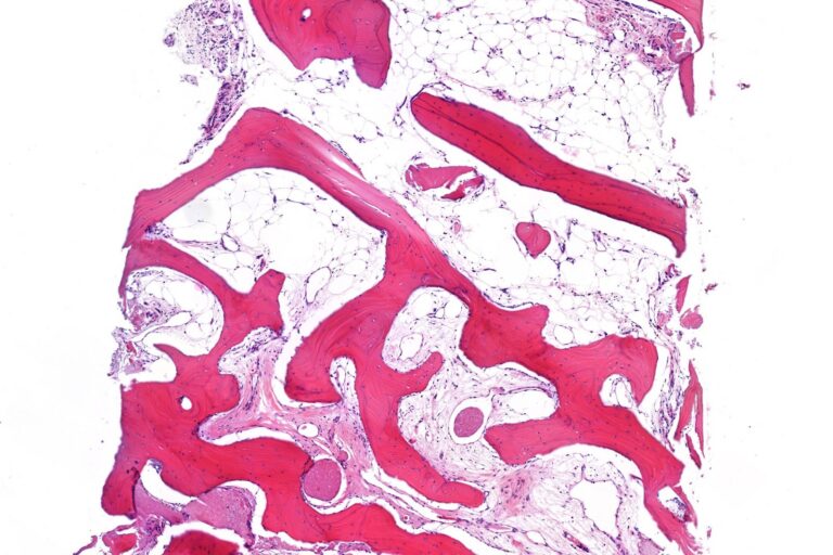
Red bone marrow produces B cells and matures them. From there they go straight into the bloodstream and travel to the secondary organs looking for pathogens. T cell precursors take a longer route, from the marrow to the thymus.
The thymus sits between the lower neck and the upper chest. Inside its cortex, developing T cells rearrange their receptor genes and face positive selection, which keeps only cells able to recognise the body’s own MHC molecules. Cells that bind too weakly die. Survivors move to the medulla, where negative selection removes any that bind the body’s own antigens too strongly, a step that prevents autoimmune disorders. Under 5 percent clear both tests.
The thymus grows from birth in response to antigen exposure and is most active in the newborn and pre-adolescent years. In adolescence it begins to shrink, and fat tissue mostly replaces its supporting structure. Some T cell production carries on through adulthood. Without a thymus, whether from birth or after its loss, a person develops severe immunodeficiency and is highly prone to infection. The thymus is not where infections are fought, though, since the T cells inside have not yet become immunocompetent.
Spleen and lymph nodes
The spleen has two working parts. Its white pulp houses lymphocytes and makes antibodies. Its red pulp, staffed by macrophages, clears aged red blood cells and pathogens. The spleen also recycles some red cell components. Haemoglobin, for instance, is broken down into amino acids that get reused.
It doubles as a store. During acute blood loss the spleen contracts and releases red cells to help maintain blood volume. Up to 25 percent of the body’s lymphocytes can be held there at once. A 2009 mouse study found that the red pulp keeps in reserve half of the body’s monocytes, which turn into dendritic cells and macrophages when they move to injured tissue such as the heart. People who lose the spleen are predisposed to certain infections.
The human body has nearly 800 lymph nodes, about 300 of them in the head and neck. Clusters sit where injuries are likely, at the groin, armpits and neck, and along the blood vessels of the intestines. Lymph arrives through several afferent vessels, seeps through the node and leaves by a single efferent one. The bean shape comes from the hilum, a dent in the surface where the efferent vessel, arteries and veins pass. When a node meets a foreign antigen, its follicles expand significantly.
Mucosal tissue and the wider system
Lymphoid tissue also lines mucous surfaces. This mucosa-associated lymphoid tissue, or MALT, includes follicles in the tonsils, Peyer’s patches and adenoids. The appendix wall is heavily infiltrated with lymphocytes too.
Lymph itself starts as fluid that leaks from the blood into body tissue. It collects waste, bacteria and damaged cells and drains into the lymphatic vessels, which pass it through the nodes to be filtered. The system moves lymph by muscle contraction and empties it back into the bloodstream through the subclavian veins. Olaus Rudbeck and Thomas Bartholin each described the system independently in the 17th century. Consult your doctor for medical advice.