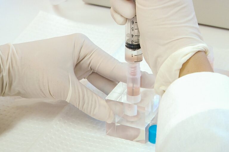
A drop of blood that can show whether a person’s brain is building the sticky clumps linked to Alzheimer’s disease: that is what two research teams reported in 2026, and for families living with memory loss it matters more than any chart.
What amyloid is, and where it comes from
Amyloid beta, written Aβ, is a small protein fragment. It is cut out of a larger molecule, the amyloid precursor protein, in what researchers call the amyloidogenic pathway. Enzymes called β-secretase and γ-secretase make the cuts. One version, Aβ42, is the troublemaker: it clumps more readily than Aβ40 and is more toxic to nerve cells.
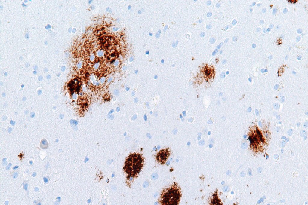
The plaques found in the hippocampus pull in more Aβ and form insoluble aggregates. They also damage mitochondria, the cell’s power units, and disturb the balance inside neurons. Immune cells called microglia and astrocytes respond with inflammation. Aβ can also switch on an enzyme that leads to abnormal tau, another protein involved in the disease.
The idea that Aβ drives all this, the amyloid hypothesis, goes back to John Hardy and David Allsop, who proposed it in 1991. Plaques themselves were noticed far earlier, in 1892.
The seeding test
Doctors have long used spinal fluid or PET scans to confirm amyloid. Both are invasive, costly and often available only at specialist centers. A group at Xuanwu Hospital, part of Capital Medical University, led by Jianping Jia, tried something different. Instead of counting how much Aβ is in blood, their assay measures how strongly the blood’s misfolded amyloid can push normal Aβ42 to aggregate, using ultrasound to amplify the effect.
“Compared to traditional quaking-based methods, this ultrasonic technology can detect Aβ oligomers at concentrations as low as 1 femtomole,” Jia said. The study ran in two stages and involved 549 participants. When the team checked it on a separate set of 429 people, the test told Alzheimer’s dementia, plus the early memory impairment it causes, apart from comparison groups. Its area-under-the-curve values ran from 0.90 to 0.93. The Alzheimer’s dementia group averaged 53.31% fluorescence, against 36.91% for cognitively unimpaired participants. The mild cognitive impairment group came in at 52.61%, so the signal was there before dementia set in.
There are limits. Every participant was of Chinese ethnicity, the design could not show who would decline later, and the work did not compare the assay with other blood markers such as p-tau217.
A second blood test
Another study, in the journal Alzheimer’s & Dementia in June 2026, looked at the ratio of plasma p-tau217 to Aβ1-42 in 499 adults aged 52 to 93 who had cognitive symptoms. The test caught nearly 98% of patients who had amyloid pathology, and it correctly cleared about 91% of those who did not.
What this means for patients is practical. Either approach, if confirmed in larger and more varied groups, could make an early answer easier to get. For now both are research findings, so anyone worried about memory should talk to a doctor.
Sources
- New blood test measures beta-amyloid seeding to identify Alzheimer's disease (The Brighter Side of News via Yahoo)
- Blood Test Accurately Detects Alzheimer's Amyloid Pathology in Symptomatic Adults (Medical Dialogues)
- New Insights Into the Pathogenesis of Alzheimer's Disease (Frontiers in Neurology)