
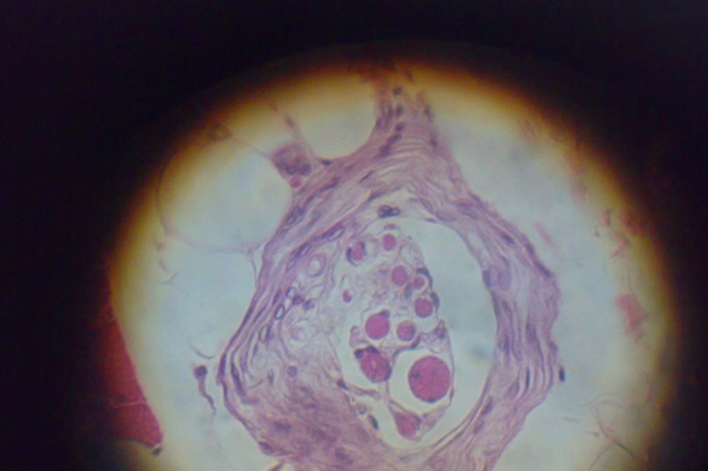
How Stretch Receptors Work Throughout the Body
Stretch receptors form a broad class of mechanoreceptors that respond to the distention of various organs and muscles across the body. They are neurologically linked to the medulla via afferent nerve fibers, which carry signals toward the central nervous system. Different types of stretch receptors serve distinct functions. The Golgi organ monitors tension in tendons, while the muscle spindle, a sensory receptor within the belly of a muscle, primarily detects changes in muscle length. Pulmonary stretch receptors are mechanoreceptors found in the lungs, and chordotonal organs perform similar roles in insects.
Beyond the digestive and muscular systems, the body relies on stretch receptors located around the carotid artery. These receptors monitor blood pressure and, when stimulated, trigger the release of antidiuretic hormone (ADH) from the posterior pituitary gland. Other examples of stretch receptors include those in the heart, the colon wall, and the arm and leg muscles and tendons. Each type is specialized for its location, but all function by detecting physical stretch or distention and converting that mechanical information into neural signals.
Gastric Receptors and the Sensation of Fullness
Among these diverse mechanoreceptors, gastric stretch receptors play a specific and crucial role in satiety. Situated in the stomach wall, they detect how much the stomach has expanded during a meal. When the stomach wall stretches, the receptors fire, and the medulla processes that information as part of the feeling of being full. The medulla, located in the brain stem, receives this input through afferent nerve fibers. This pathway helps the brain interpret the degree of stomach expansion, contributing directly to the perception of fullness.
The process does not begin with the stomach as a whole but with these specialized receptors responding to physical distention. The signals travel to the medulla, which integrates them with other sensory and metabolic cues to produce the sensation that tells a person to stop eating. This mechanism is part of a larger system in which the brain manages complex responses to internal stimuli, ensuring that the body maintains energy balance.
The Brain’s Role in Integrating Sensory Information
The brain is the central organ of the nervous system in vertebrates and most invertebrates, responsible for processing sensory information, coordinating movement, and regulating bodily functions. In vertebrates, it develops from the dorsal nerve cord and is divided into the forebrain, midbrain, and hindbrain, with the spinal cord extending its control throughout the body. This centralized structure allows the brain to manage complex responses to both internal and external stimuli.
Stretch receptors are part of this system, detecting distention in organs and muscles and relaying signals to the medulla in the brain stem. The brain is the organ that receives and interprets these signals, enabling functions such as satiety, blood pressure regulation, and muscle coordination. Understanding how the brain processes such signals helps explain fundamental bodily sensations, including the feeling of fullness after eating. This context highlights the brain’s role in integrating information from stretch receptors to maintain homeostasis and guide behavior. By deciphering these neural pathways, researchers gain insight into how the body senses its own internal state and adjusts behavior accordingly, a key aspect of maintaining health.