
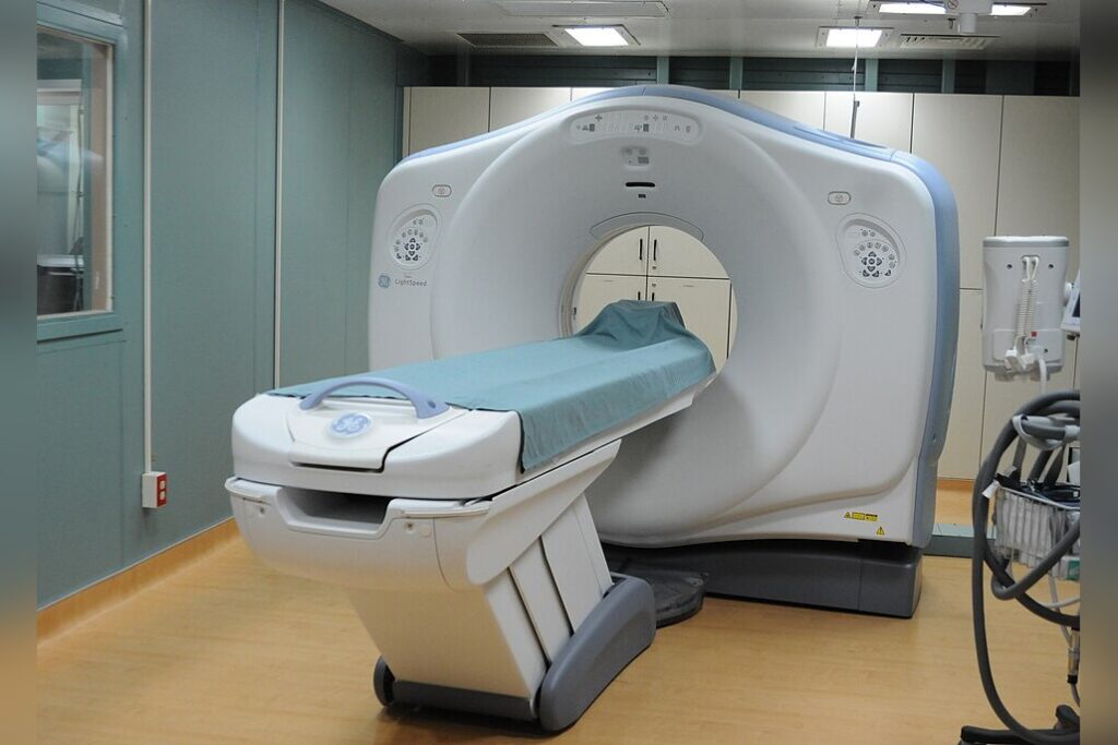
A single computed tomography scan failed to detect a brain lesion in Tyler Morton. That lesion later appeared on a second CT scan, but by then the tumor had already reached an advanced stage. Morton’s experience highlights a critical gap in diagnostic imaging: CT scans, while fast and widely used in emergency settings, have well-documented blind spots that can delay the detection of certain brain tumors.
The Limits of a First Look
CT scans are frequently employed in emergency departments because they are rapid and readily available for assessing acute neurological symptoms. However, the technology has inherent limitations. According to the American Cancer Society, CT scans can miss small or low-grade tumors, particularly those located in the brainstem or posterior fossa areas of the skull.
This lower sensitivity for brain tumors compared to other imaging methods means that some lesions simply are not visible on a CT scan until they grow larger. In Morton’s case, the first scan produced no evidence of a lesion, while the second scan–taken later–did reveal the tumor, but by that point it was already advanced.
The delay in diagnosis may have been influenced by a reliance on CT scanning alone, raising questions about when a faster but less sensitive tool is sufficient.
Guidelines and the Case for MRI
Magnetic resonance imaging offers a fundamentally different approach. MRI provides superior soft-tissue contrast, making it the preferred imaging modality for suspected brain tumors.
The National Institute for Health and Care Excellence, known as NICE, recommends urgent MRI for patients with progressive neurological symptoms. That guideline exists precisely because of situations like Morton’s: when a CT scan misses what an MRI would catch. MRI technology developed during the 1970s and 1980s, growing out of nuclear magnetic resonance.
The word “nuclear” was eventually dropped from the name to avoid negative associations. The process works by using strong magnetic fields and radio waves to excite hydrogen atoms–abundant in human water and fat–and antennas detect the radiation emitted to map internal structures.
Unlike CT scans, MRI does not use X-rays or ionizing radiation. But MRI scans take longer and are noisier than CT scans, and some patients cannot undergo MRI because metal implants in the body pose safety risks. Despite these trade-offs, the higher contrast resolution makes MRI a more sensitive tool for detecting small or subtle brain abnormalities.
Balancing Speed and Sensitivity
Morton’s case is a single data point, but it points to a broader issue in neurological diagnostics: the trade-off between speed and sensitivity. CT scans are invaluable for rapid assessment in emergencies, but their blind spots can lead to missed diagnoses when a tumor is small or located in an area where CT sensitivity is reduced. Using the more detailed images provided by MRI can lead to earlier detection of abnormalities in the brain. For patients with progressive neurological symptoms–symptoms that worsen over time–the guidelines recommend MRI as the appropriate tool.
The stakes are high, as demonstrated by Morton’s experience. A clean CT scan does not always mean a clean bill of health.
Choosing the right imaging tool affects how quickly doctors find a disease, and that timing can influence treatment options and outcomes. While no single case dictates universal policy, the discrepancy between Morton’s first scan and the later finding of an advanced tumor underscores the need for clinicians to consider MRI when symptoms persist, even after a normal CT result. (Patients should consult their doctor for medical advice regarding their specific situations.)
{“@context”: “https://schema.org”, “@type”: “NewsArticle”, “headline”: “CT Scans Miss Early Brain Tumors, Highlighting Critical Gaps”, “description”: “WASHINGTON, July 17 — A single computed tomography scan failed to detect a brain lesion in Tyler Morton. That lesion later appeared on a second…”, “datePublished”: “2026-07-17T09:30:00+00:00”, “author”: {“@type”: “Person”, “name”: “Li Wei”}, “publisher”: {“@type”: “Organization”, “name”: “InfoPulse Today”, “url”: “https://newsfeed.infopulsetoday.com/”}}